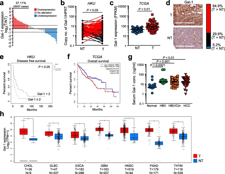
Fig. 1.
Overexpression of Gal-1 was associated with poor clinical outcome in HCC patients. a. Gal-1 overexpression in 37.11% (36/97) of paired HCC cases comparing tumorous (T) and non-tumorous (NT) tissues. b. HKU cohort consisted of a significant overexpression of Gal-1 (n = 97). c. This trend was similarly observed in The Cancer Genome Atlas (TCGA) cohort for liver cancer (n = 50). d. Intense Gal-1 staining in tumorous tissues compared to non-tumorous tissues found in 64.9% of cases through IHC analysis. e. Clinicopathological analysis of patients with Gal-1 overexpression indicated poorer disease-free survival. f. Based on TCGA database, Gal-1 overexpression was found to be associated with poorer overall survival. g. The level of Gal-1 secretory analyzed by ELISA in patient blood serum revealed the increased Gal-1 levels in patients with HBV, cirrhosis and HCC. HBV: hepatitis B virus; Cirr: cirrhosis; HCC: hepatocellular carcinoma. h. Identification of Gal-1 overexpression observed in other cancers from TCGA database comparing tumor and non-tumor counterparts. CHOL: cholangioma; DLBCL: diffuse large B-cell carcinoma; ESCA: esophageal carcinoma; GBM: glioblastoma; HNSC: head and neck squamous cell carcinoma; PAAD: pancreas adenocarcinoma; THYM: thymoma. *P < 0.05 is considered to be statistically significant

