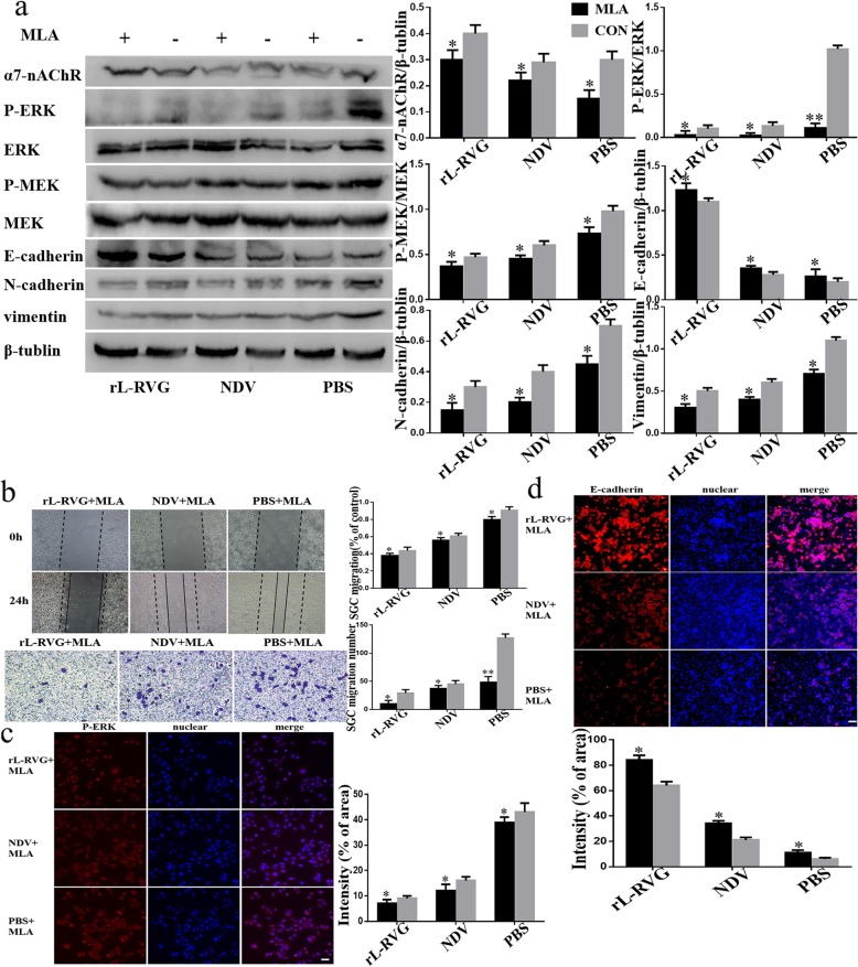
Fig. 4.
Effects of rL-RVG and MLA pretreated cells on α7-nAChR, MEK/ERK signaling pathway and epithelial/mesenchymal proteins and migration of cells. a Western blot analysis of the α7-nAChR, MEK/ERK signaling pathway and epithelial/mesenchymal protein markers. b Cell migration was detected by wound healing and transwell assay. c Immunofluorescence analysis of P-ERK. d Immunofluorescence analysis of EMT markers E-cadherin in infected SGC cells. *P < 0.5,**P < 0.01 (rL-RVG + MLA vs rL-RVG, NDV + MLA vs NDV, PBS + MLA vs PBS,Bar = 25 μm)

