Fig. 5.
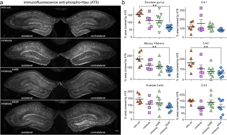
Tau-degrading intrabodies eliminate tauopathy at mid-disease in P301S-tg mice. a. Representative images of pathological tau accumulation as seen with AT8 staining in 9.5 month-old P301S-tg mice following the expression of anti-tau intrabodies after tauopathy deposition (mid-disease). b AAV-control displayed an increase in pathological tau accumulation in the ipsilateral hippocampal side. Expression of the conventional anti-tau intrabody displayed a non-significant increase in pathological tau accumulation in the dentate gyrus, granule cells, mossy fibers and CA2 ipsilateral relative to the contralateral hippocampus. Expression of the chimeric tau-degrading intrabody fused to ubiquitin harboring a K48R mutation prone for lysosomal-meditated degradation displayed a non-significant increase in pathological tau accumulation in the granule cells, CA1 and CA3 regions of the ipsilateral relative to the contralateral hippocapus. Expression of the chimeric tau-degrading intrabody fused to ubiquitin harboring a K63R mutation prone for proteasome-mediated degradation displayed a significant decrease in pathological tau accumulation in the hippocampal dentate gyrus, granule cells and mossy fibers in the ipsilateral relative to the contralateral hippocampus. Scale bar 200 μM. Quantification (n = 6–9) of pathological tau from the contralateral to the ipsilateral dentate gyrus. All data are mean ± s.e.m. one-way ANOVA with Tukey’s Multiple Comparison. *p < 0.05, **p < 0.01

