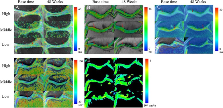
Fig. 4.
Representative images of quantitative MRI mappings of three patients from high (60 years, F), middle (68 years, M), and low (65 years, M) dose groups. For the same patient, the change (red arrow) of the T1rho (a) image was more obvious than that of the T2 (b), T2star (c), R2 star (d), and ADC (e) images. Changes of T1rho values between two examination time points in the patient from the high-dose group were more pronounced than those from the other two groups, whereas this phenomenon was not found in other MRI images

