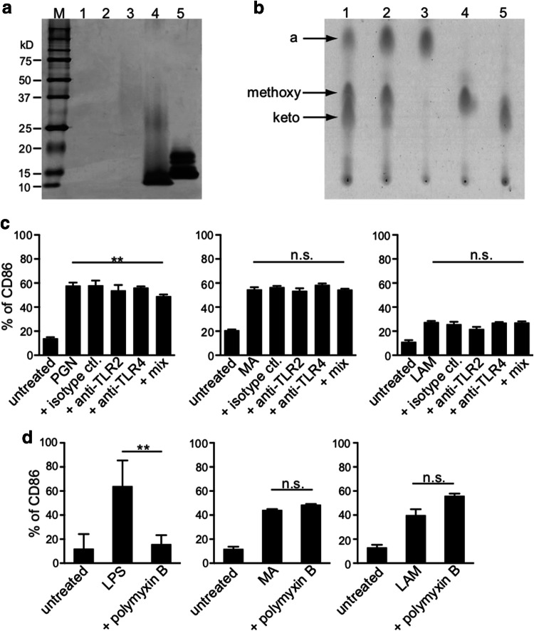
Fig. 2.
Confirmation of MA and LAM purity. a Protein detection by silver stain SDS-PAGE gel. Lane 1: 10 µg MA from BCG isolate, Lane 2: 10 µg MA from Aoyama B isolate, Lane 3: 10 µg LAM from Aoyama B isolate, Lane 4: 1 µg PGN from Staphylococcus aureus, Lane 5: 1 µg LPS from Escherichia coli. Numbers shown in the left-axis indicate molecular weights (kD). b Thin-layer chromatography of purified MA. Lane 1: MA from BCG, Lane 2: MA from Aoyama B isolate, Lane 3: α-MA from Aoyama B isolate, Lane 4: methoxy-MA from Aoyama B isolate, Lane 5: keto-MA from Aoyama B isolate. All samples were 50 µg. c CD86 expression on MDDCs stimulated by 10 µg/mL PGN (left), 200 µg/mL MA from Aoyama B isolate (middle), 200 µg/mL LAM (right) treated with anti-TLR2/4 antibodies. Data are shown as the mean ± SD of n = 4 samples per measurement. d CD86 expression on MDDCs stimulated by 100 ng/mL LPS (left), 200 µg/mL MA from Aoyama B isolate (middle), 200 µg/mL LAM (right) with or without 15 µg/mL polymyxin B. Data are shown as the mean ± SD of n = 4 samples per measurement. **p < 0.01, n.s. not significant; one-way ANOVA followed by Bonferroni post-tests

