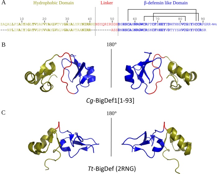
FIG 8.
Cg-BigDef1[1–93] and Tt-BigDef 3D structure comparison. The hydrophobic domain, the linker, and the β-defensin-like domain are colored in deep olive, red, and blue, respectively. (A) Alignment of Cg-BigDef1[1–93] (top) and Tt-BigDef (bottom) primary sequences. Conserved residues are indicated in bold. (B) 3D structure (cartoon representation) of Cg-BigDef1[1–93] (PDB entry 6QBL). (C) 3D structure (cartoon representation) of Tt-BigDef (PDB entry 2RNG).

