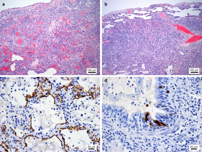
FIG 2.
Histopathology of oseltamivir-treated animals. (a and b) Hematoxylin and eosin (HE) staining of the lungs of oseltamivir-pretreated (a) and -posttreated (b) surviving animals at day 20 postinfection, showing a chronic healing stage of pneumonia with areas of consolidation and thickening of alveolar walls due to infiltration of mononuclear cells and deposition of fibrous tissue. (c and d) The presence of viral antigen was demonstrated using an antibody directed to the viral nucleoprotein and was detectable only at the time of euthanasia in the controls (not shown) and animal POST1, which was euthanized on 9 dpi. Viral nucleoprotein was found in alveolar cells (c) and bronchiolar epithelium (d).

