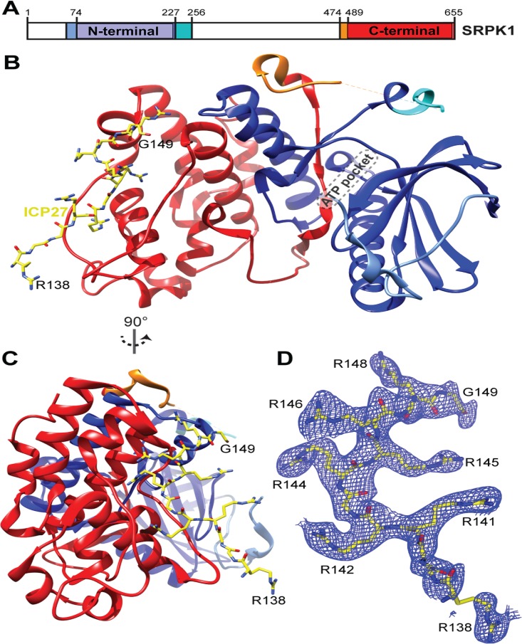
FIG 3.
Structure of SRPK1 with RGG box peptide ICP27137–152 bound in the substrate docking groove. (A) Schematic of SRPK1 domains. (B) Crystal structure of the SRPK1 in complex with ICP27137–152. SRPK1 is colored as panel A, ICP27 is yellow. (C) As in panel A but view rotated 90°. (D) 2Fo − Fc electron density map of RGG peptide colored blue, contoured to 1σ orientated as in panel B.

