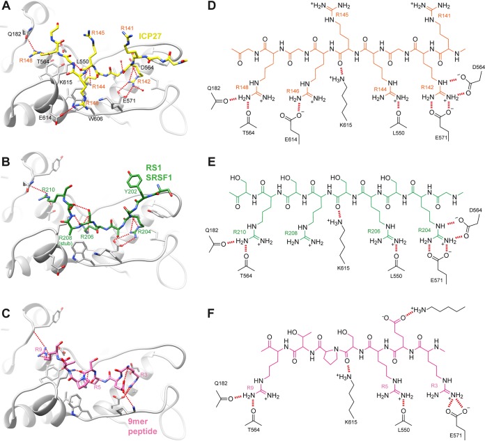
FIG 4.
Comparison of SRPK1 docking groove interactions. (A) Detail of the RGG box peptide bound within the SRPK1 docking groove, plus comparative views of SRPK1 substrate complexes: (B) SRSF1 RS1 region (8) or (C) 9-mer docking peptide of membrane-associated guanylate kinase (7). SRPK1 is shaded gray, and bound peptides are colored. Intermolecular hydrogen bonds or salt bridges are represented by red dashes. Chemical schematics of the interactions shown in panels A to C are shown in panels D to F, respectively.

