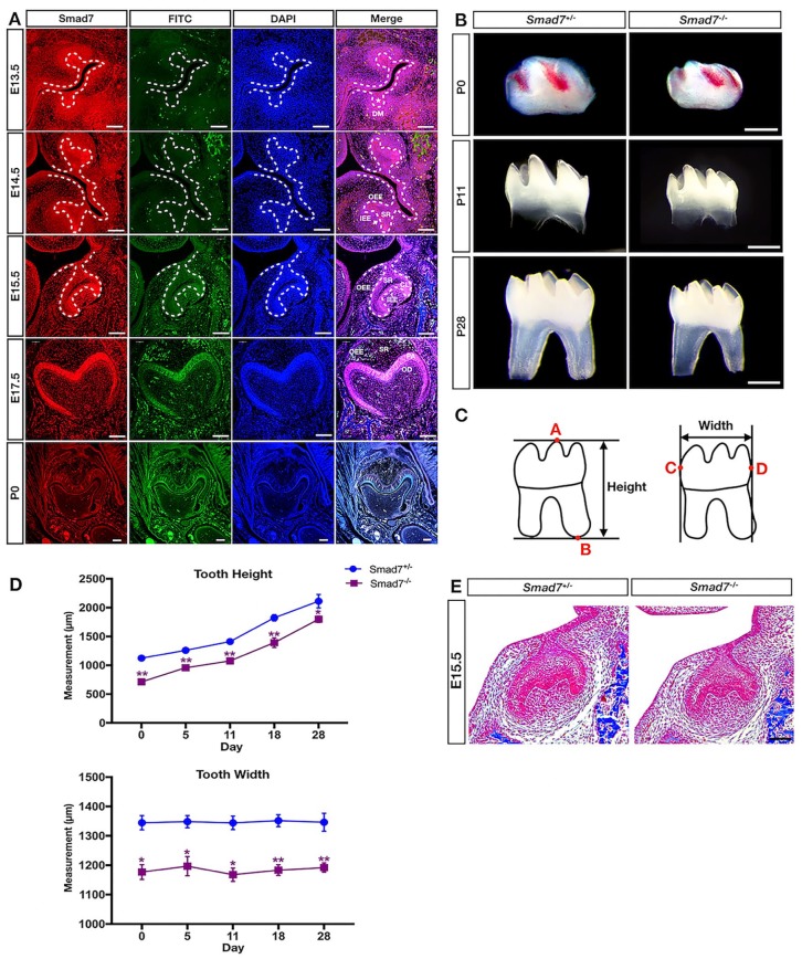
Figure 1.
Smad7 deficiency leads to reduced tooth size. (A) Immunofluorescent staining of Smad7 during tooth morphogenesis at the bud stage (E13.5), cap stage (E14.5), early bell stage (E15.5), late bell stage (E17.5), and postnatal stage (P0). (B) Representative images of control and Smad7−/− lower first molar at P0, P11, and P28 show unaltered dental patterning with reduced size in the mutants. (C) Schematic representation of the methods used for measuring the molar size, including height and width. Point A: the tip of the highest cusp of the molar; point B: the bottom of the molar root; point C: the most convex point on the mesial surface of the tooth crown; point D: the most convex point on the distal surface of the tooth crown. (D) Measurements of tooth size of the control and Smad7−/− first mandibular molars at different postnatal time points (n = 4 for each time point). Statistical analysis was performed using Student’s t test. *P < 0.05. **P < 0.01. (E) Representative histology from the control and Smad7−/− first mandibular molars at E15.5 shows discernably reduced size in the mutant. CL, cervical loop; DE, dental epithelium (marked by asterisk); DM, dental mesenchyme; IEE, inner enamel epithelium; OEE, outer enamel epithelium; PEK, primary enamel knot (indicated by arrow); SR, stellate reticulum. Scale bars: 500 µm (B); 100 µm (A and E).

