Figure 2.
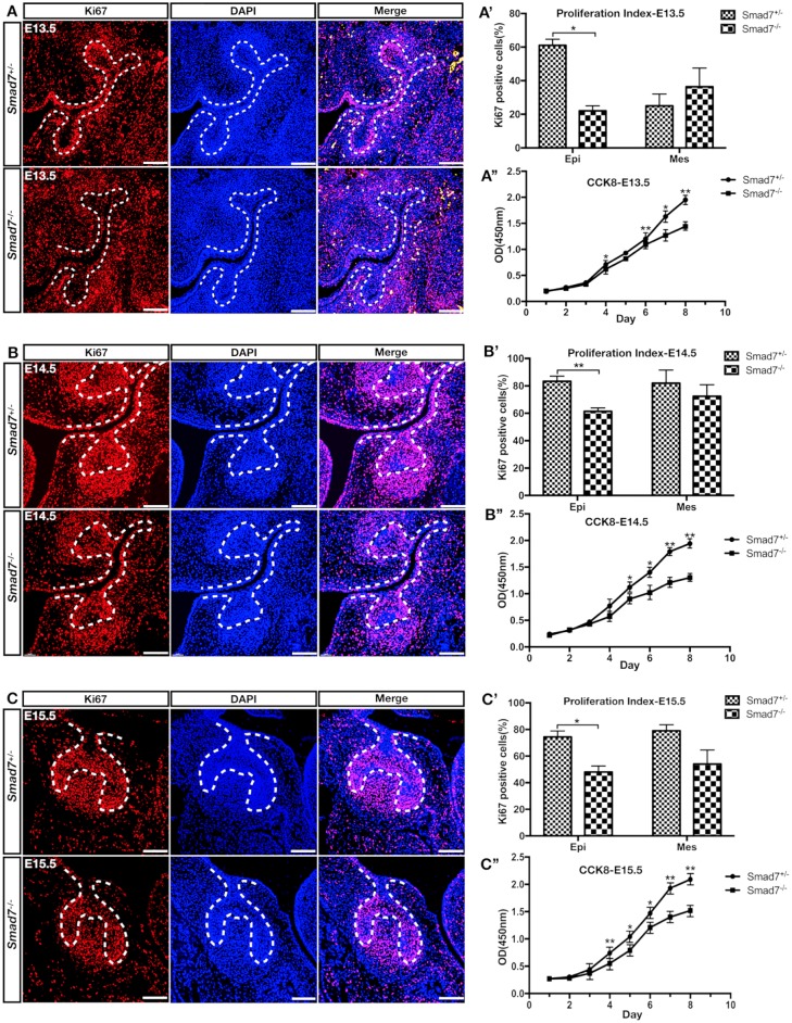
Inactivation of Smad7 leads to compromised cell proliferation in the developing molar. (A–C) Immunofluorescent staining of Ki-67 on sections of control and Smad7−/− molar germs at E13.5 (A), E14.5 (B), and E15.5 (C). (A′–C′) Quantification of Ki-67–positive cells in the dental epithelium and mesenchyme of control and Smad7−/− tooth germs at E13.5 (A′), E14.5 (B′), and E15.5 (C′). (A′′–C′′) Growth curves of control and Smad7−/− tooth germ cells plotted from CCK8 assays at E13.5 (A′′), E14.5 (B′′), and E15.5 (C′′). Statistical analysis was performed using Student’s t test. *P < 0.05. **P < 0.01. Scale bars: 100 µm.

