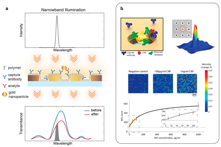
Figure 7.
(a) Nanoparticle-enhanced plasmonic imaging. Schematic shows a collinear transmission light-path, where a narrowband illumination tuned to the flank of the Au nanohole array resonance peak is used for bright-field imaging. Nanoparticle binding at the nanoholes distort the localized plasmons creating a drastic suppression of the transmission peak, which can be monitored to extract digital analyte binding information; (b) (top) schematic shows a sandwich bioassay, antigen being recognized by capture antibodies immobilized on the Au-nanoholes and then by detection antibodies tethered to Au-nanoparticles. Strong local suppression in the transmission create intensity contrast. (bottom) Bright-field images and a calibration curve for human C-reactive Protein detection. Adapted with permission from [90] Copyright 2018 American Chemical Society.

