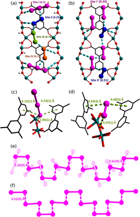
Figure 3.

Views along the b axis of I2-loaded (a) MFM-300(VIII) and (b) MFM-300(VIV) obtained by high-resolution synchrotron PXRD. Views of binding sites for I3– and I2 in (c) MFM-300(VIII) and (d) MFM-300(VIV), respectively. Views of I2 (solid) and I3– (pale wire frame) in (e) I2@MFM-300(VIII/IV) and (f) I2@MFM-300(VIV).
