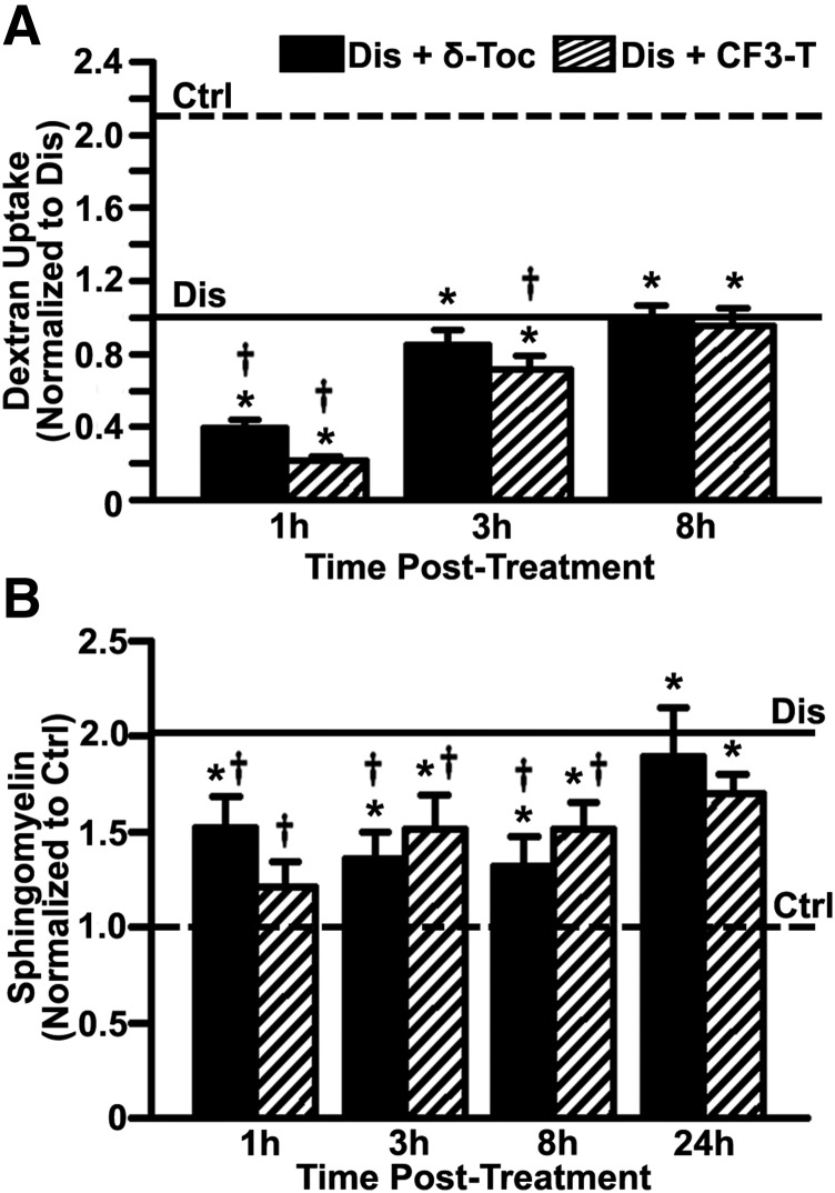
Fig. 8.
Kinetics of tocopherol effect on bulk fluid-phase uptake and sphingomyelin levels. (A) Control (Ctrl) and imipramine-diseased endothelial cells (Dis) were incubated with Texas Red dextran (1 hour pulse) 1 to 8 hours after a 48-hour treatment with 40 µM δ-tocopherol or 20 µM CF3-T. Dextran uptake was normalized to untreated diseased cells (horizontal solid line). Dextran uptake by untreated control cells is shown by the horizontal dashed line. (B) Sphingomyelin was stained with fluorescent lysenin 1 to 24 hours after a 48-hour treatment with δ-tocopherol or CF3-T, quantified as described in Fig. 1, and normalized to untreated control cells (horizontal dashed line). Sphingomyelin levels in untreated diseased cells are shown by the horizontal solid line. Data are mean ± S.E.M. (n ≥ 4 independent wells). *Comparison with untreated control cells; †comparison with untreated diseased cells (P < 0.05 by Student’s t test).

