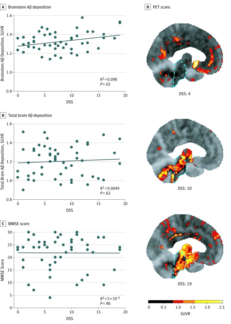
Figure 1. Associations of Daytime Sleepiness Score (DSS) With β-Amyloid (Aβ) Deposition and Mini-Mental State Examination (MMSE) Performance.
A-C, The DSS was associated with brainstem Aβ deposition (B = 0.0063; 95% CI, 0.001 to 0.012; P = .02) (A), but not with total brain Aβ deposition (B = 0.0023; 95% CI, −0.007 to 0.011; P = .62) (B) or MMSE scores (B = −0.01; 95% CI, −0.39 to 0.37; P = .96) (C). Data points represent individual patients. D, Representative fluorine 18–labeled florbetaben Aβ positron emission tomography (PET) scan images from 3 different patients with DSSs of 4, 10, and 19, respectively, are shown. A DSS of 10 or greater indicates excessive daytime sleepiness. The location of the brainstem in each scan is highlighted by a dashed cyan outline. Scale bar indicates units of standardized uptake value ratio (SUVR), with higher SUVRs indicating higher levels of Aβ deposition.

