Figure 2.

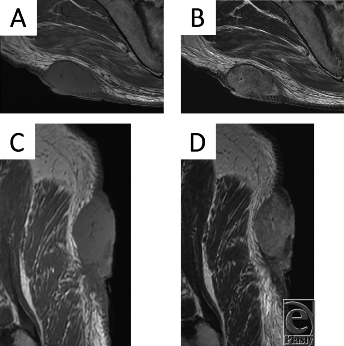
(a) Axial T1-weighted MRI scan of the tumor. (b) Axial T2-weighted MRI scan of the tumor. (c) Sagittal T1-weighted MRI scan of the tumor. (d) Sagittal T2-weighted MRI scan of the tumor.

(a) Axial T1-weighted MRI scan of the tumor. (b) Axial T2-weighted MRI scan of the tumor. (c) Sagittal T1-weighted MRI scan of the tumor. (d) Sagittal T2-weighted MRI scan of the tumor.