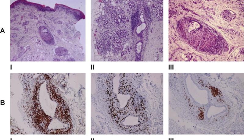
Fig. 2.

Histopathological findings in the labial mucosa. A – I. The biopsy of the swollen lip shows granulomatous chelitis characterized by perivascular lymphocytic infiltration with granuloma formation in the submucosa (haematoxylin-eosin [H&E], original magnification x 40). II. Deeper areas of the biopsy show the presence of chronic inflammatory infiltration of the lips' salivary glands (H&E, original magnification x 100). III. High power view of the granuloma (H&E, original magnification x 100). B – Immunochemistry study. I. CD4+ cells representing 75% of T-Lymphocytes. II. CD8+ cells representing 15% of T-Lymphocytes. III. Scanty CD20+ cells (x100 immunoperoxidase with haematoxylin counterstain).
