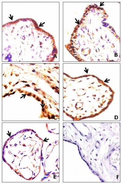
Figure 1.

(A) Control placenta (40 weeks) showing moderate cytoplasmic expression of HIF‐1α in syncytiotrophoblast; (B and C) preeclamptic placenta (38 and 36 weeks) showing nuclear accumulation of HIF‐1α in syncytiotrophoblast; (D) control placenta (32 weeks) showing moderate cytoplasmic expression of PIGF in syncytiotrophoblast; (E) preeclamptic placenta (32 weeks) showing mild cytoplasmic expression of PIGF in syncytiotrophoblast; (F) negative control incubated with IgG showing placental villi. Arrow shows the expression of protein. Magnification: 400×.
