
Figure 1.
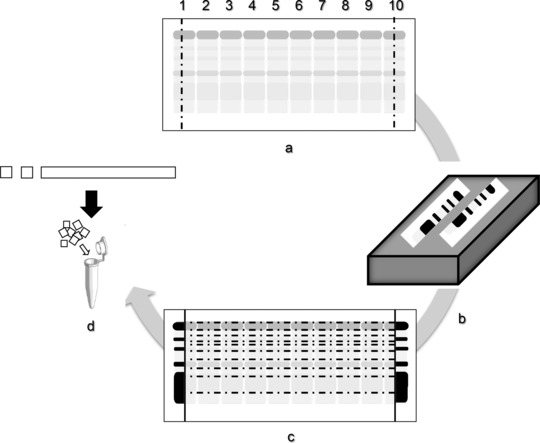
Schematic representation of the protocol for protein extraction from CAM. Loading of ten lanes with urine sample on CAM (a), silver staining of both ends after separation (b), cutting each fraction of the unstained membrane following the stained area (c), and fragmenting each fraction strip for protein extraction (d).