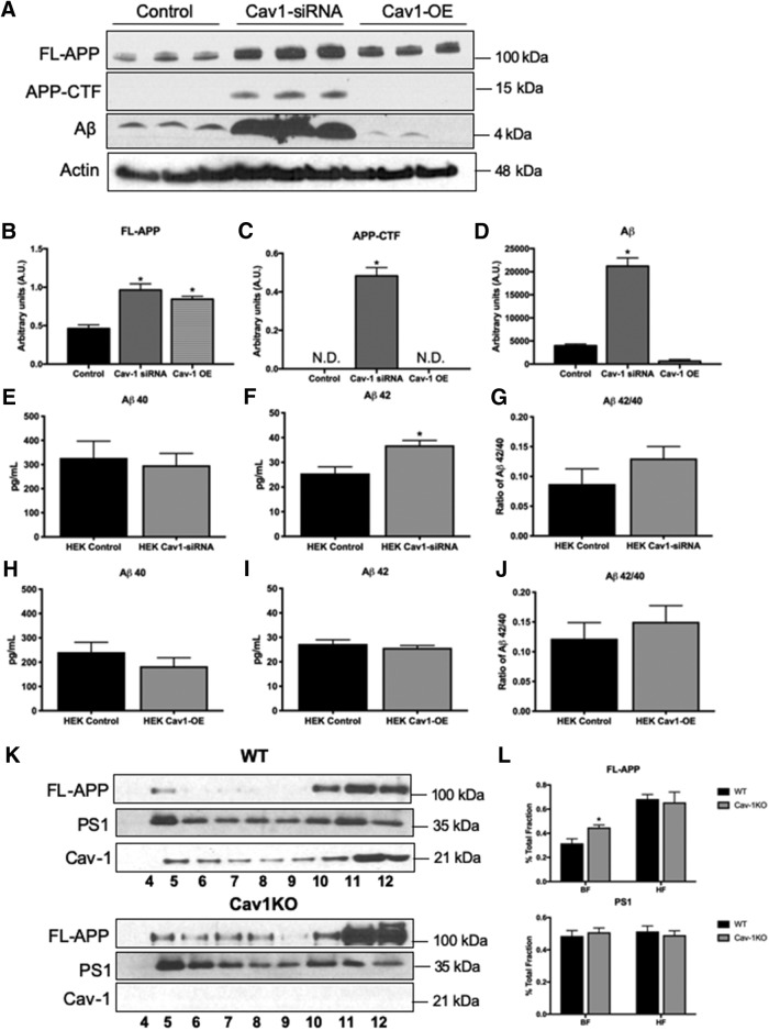
Figure 4.
Depletion of Cav-1 induces alterations in APP processing. A–D, Comparison of protein levels in HEK-APPswe cells expressing the control, Cav-1-siRNA, or Cav-1 overexpressing virus (Cav1-OE). Downregulation of Cav-1 induces increases in FL-APP (A,B), APP-CTF (A,C), and Aβ (A,D), whereas overexpression of Cav-1 partially restores FL-APP levels (A,B), and fully restores the expression levels of APP-CTF (A,C) and Aβ (A,D) (N = 3, one-way ANOVA with multiple comparisons; B, F(2,6) = 19.81, p = 0.0023; C, F(2,6) = 122.7, p < 0.0001; D, F(2,6) = 107.5, p < 0.0001). E–J, Analysis of the concentration of human Aβ42 and Aβ40 secreted from HEK-APPswe cells infected with control virus, Cav-1-siRNA, or Cav-1 overexpressing virus (Cav1-OE) was performed by ELISA (N = 3). *p < 0.05 (unpaired t test). Levels of Aβ40 were unchanged (E,H), but levels of Aβ42 were significantly increased following Cav-1 downregulation (F). The ratio of Aβ42/40 shows a trending increase following Cav-1 downregulation (G; p = 0.09). Following Cav-1 upregulation, levels of Aβ42 (I) and the ratio of Aβ42/40 (J) were comparable with control. K, Western blot analysis of sucrose density fractionation probed for expression levels of FL-APP, PS1, and Cav-1. L, Quantification of sucrose density fractionation shows that Cav-1KO displays a significant increase in the expression and localization of FL-APP to the lipid raft fractions. Levels of PS1 in the lipid raft fractions are unchanged (N = 3). *p < 0.05 (unpaired t test).

