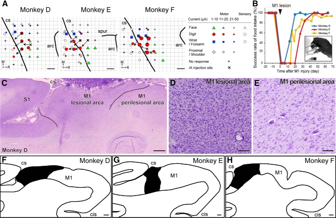
Figure 1.
Lesioning of primary M1 hand area and subsequent motor recovery. A, Injection site of ibotenic acid superimposed on ICMS maps of M1 in each lesioned monkey. Arrows indicate the orientation of the blade face at the needle tip. Movement elicited at the threshold and the sensory response to light tactile stimuli is indicated by the symbols. Some electrode penetration sites showed no response to either ICMS at current strengths ≤50 μA or sensory stimulation. B, Time course of changes in the success rate of precision grip in the lesioned monkeys. A successful trial was defined as the removal of the object by using the precision grip between the tip of the index finger and thumb, as shown in the picture. C–E, Nissl-stained section of Monkey D showing a representative lesion of M1. Ibotenic acid injection in M1 resulted in gliosis and loss of neurons (D), whereas neurons were observed in the neighboring area, defined as the perilesional area. Dotted lines indicate the boundaries between the lesional area and the perilesional area. F–H, Line drawing of the M1 hand area in each lesioned monkey. Black region indicates the lesional area. CS, Central sulcus; arc, arcuate sulcus; spur, spur of arcuate sulcus; M, medial; R, rostral; IA, ibotenic acid; S1, primary sensory area; cis-, cingulate sulcus. Scale bars: A, 2 mm; C, F–H, 1 mm; D, E, 300 μm.

