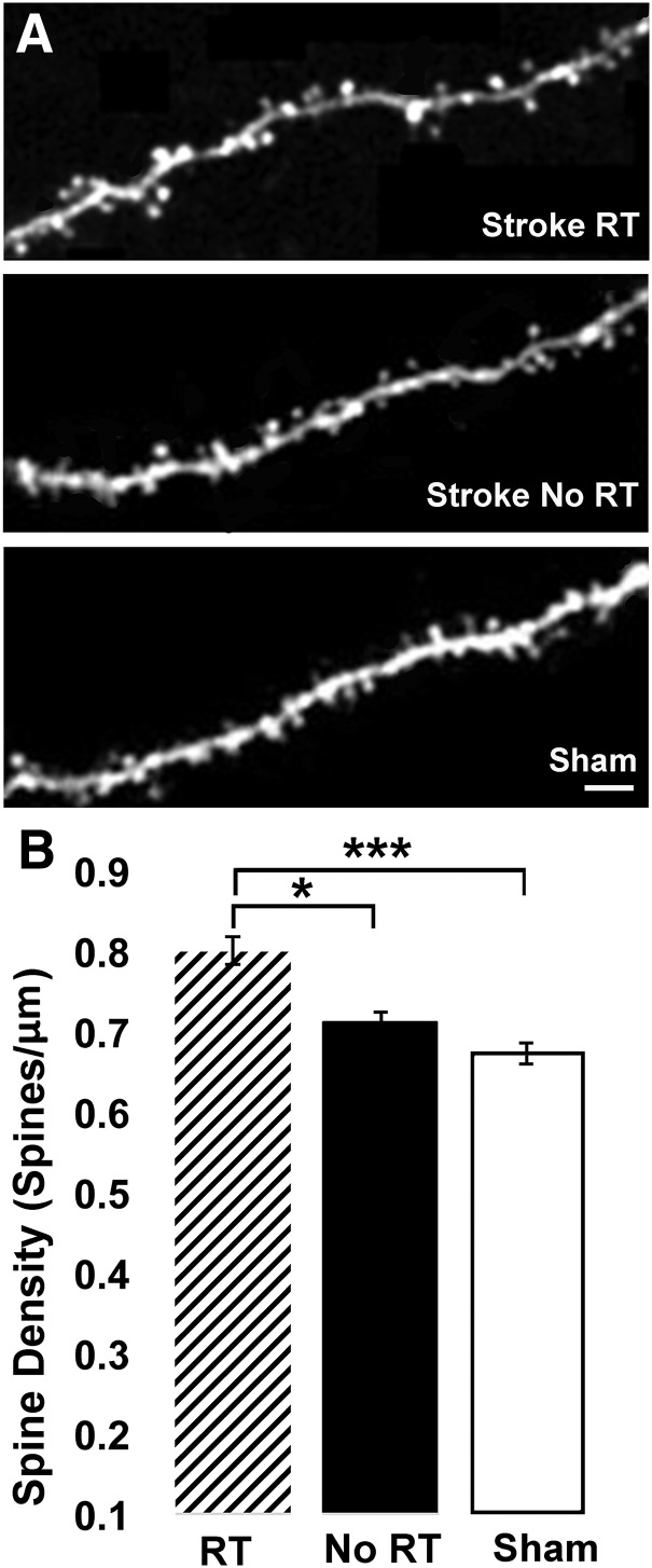
Figure 7.
RT increased spine density on apical dendrites of layer V pyramidal neurons in layer II/III of peri-infarct MC. A, Confocal image of an apical dendrite sampled in layer II/III from the RT (top), No RT (middle), and Sham (bottom). Scale bar, 10 μm. B, Spine density, measured as spines/μm, was increased in the RT group at 8 weeks after the infarct relative to both No RT (*p < 0.05) and Sham (***p < 0.0001). Spine density was not significantly increased in the No RT group compared with Sham (p = 0.07).

