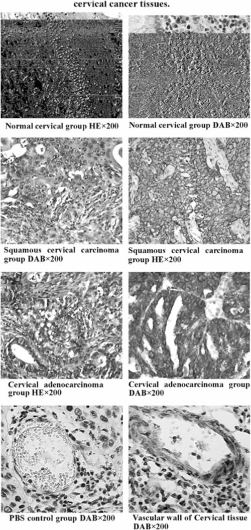
Figure 1.

Immunohistochemical staining (SABC method) results of cervical cancer tissues. (1) Normal cervical group HE × 200. (2) Normal cervical group DAB × 200. (3) Squamous cervical carcinoma. (4) Squamous cervical carcinoma group DAB × 200 group HE × 200. (ref SHAPE MERGEFORMAT ref SHAPE MERGEFORMAT 5) Cervical adenocarcinoma. (6) Cervical adenocarcinoma group HE × 200 DAB × 200. (ref SHAPE MERGEFORMAT 7) PBS control group DAB × 200. (8) Vascular wall of Cervical tissue DAB × 200.
