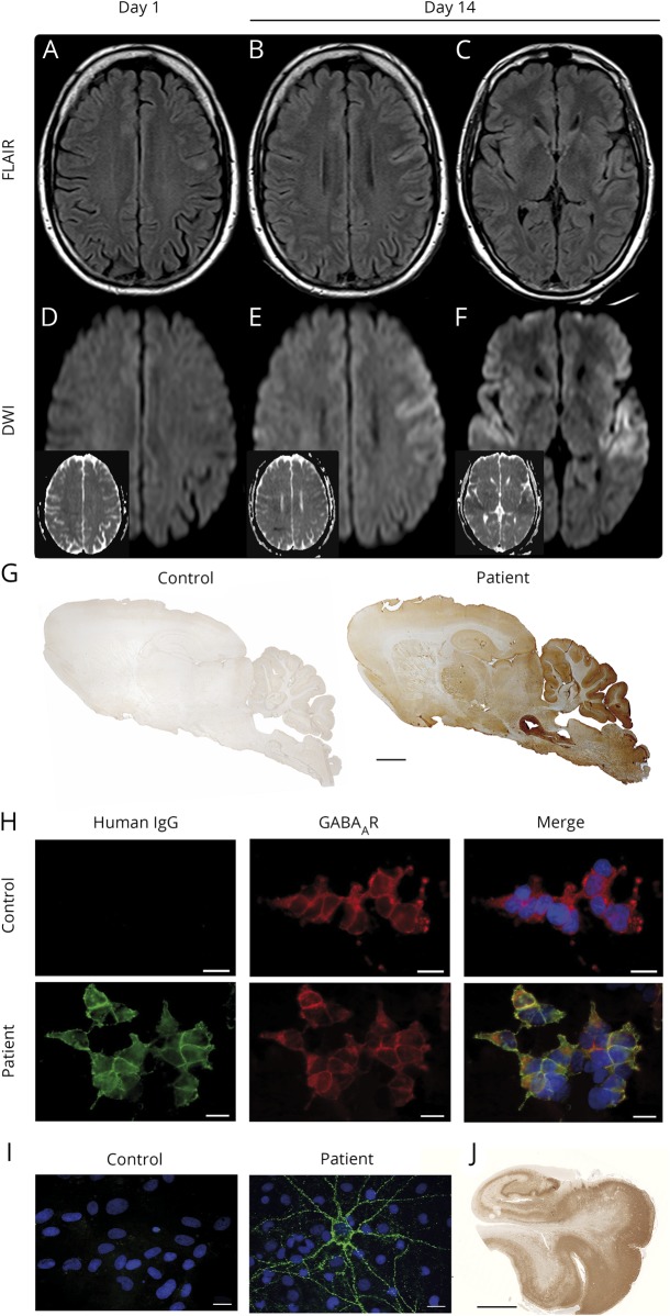
Figure. MRI findings and laboratory studies.
MRI 1st day of admission (A and D) and 14 days after admission (B, C, E, and F), showing progression of the left frontal hyperintense lesion and new diffusion restriction left frontally and bilaterally in the operculum on day 14. (A–C) Axial fluid-attenuated inversion recovery-weighted images with hyperintense lesion of the left prefrontal gyrus (A and B) and the operculum bilaterally (C). (D–F) Diffusion-weighted images and apparent diffusion coefficient images (small insets) without diffusion restriction (D) and marked diffusion restriction in the left prefrontal gyrus (E) and the operculum bilaterally (F). (G) Immunolabeling of sagittal rat brain sections with the patient's CSF antibodies showing a characteristic pattern. Patient and control CSF 1:4. Anti-human IgG (H + L). Human IgM and IgA did not show immunoreactivity. Scale bar 1 mm. (H) Detection of antibodies to the GABAA receptors (GABAAR) using HEK293 cell-based assay. Patient's but not control serum detects GABAAR. Human GABAAR subunits transfected into HEK293 cells and stained via life cell staining (serum 1:40). Green human IgG, red commercial GABAAR antibody. Scale bar 20 μm. (I) Patient's but not control serum detects neuronal surface antigens. Nonpermeabilized embryonic rat hippocampal neuron cultures DIV21 life cell stained with human IgG and nuclear counterstaining with DAPI (blue). Scale bar 20 μm. (J) Postmortem herpes simplex virus antigen staining of the patient's hippocampus. Scale bar 5 mm. DAPI = 4′,6-diamidino-2-phenylindole; IgG = immunoglobulin G.

