
Figure 3.
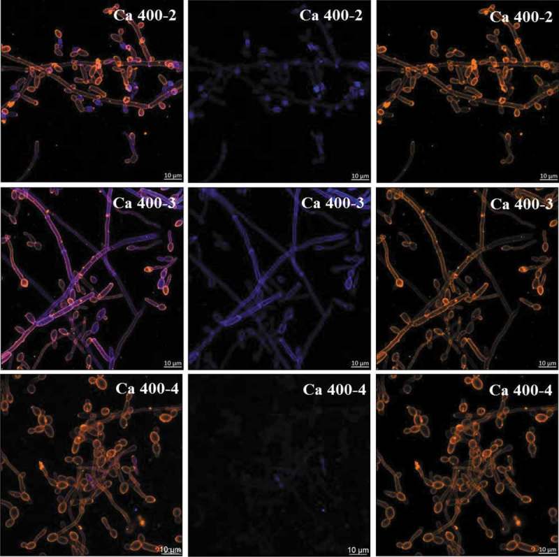
Representative confocal microscopy images of 19 h-old C. albicans single-species biofilms. These biofilms are labeled with primary antibody (400–2, 400–3, 400–4) paired with secondary antibody labeled with Alexa Fluor 405 (shown in blue). The orange color represents microbial cells of the biofilm labeled by lectin concanavalin A conjugated with TRITC. The blue color represents polysaccharides produced by C. albicans. In the first column the overlay is observed, while the second and third illustrate each component individually.