
Figure 4.
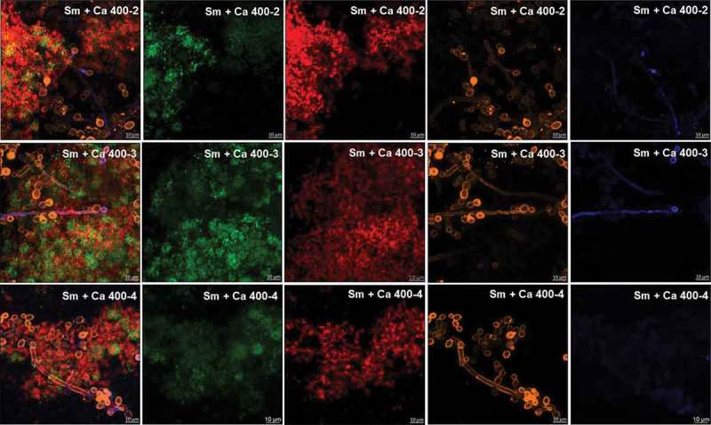
Representative confocal microscopy images of 19 h-old S. mutans and C. albicans dual-species biofilms. The green and orange colors represent cells of S. mutans (GFP) and C. albicans (concanavalin A conjugated with TRITC), respectively. The red color represents exopolysaccharides produced by S. mutans (Alexa Fluor 647) and the blue color represents polysaccharides produced by C. albicans (labeled with primary antibody 400–2, 400–3 or 400–4, and paired with secondary antibody conjugated to Alexa Fluor 405). The overlay is observed in the first column, while the other columns represent each component individually.