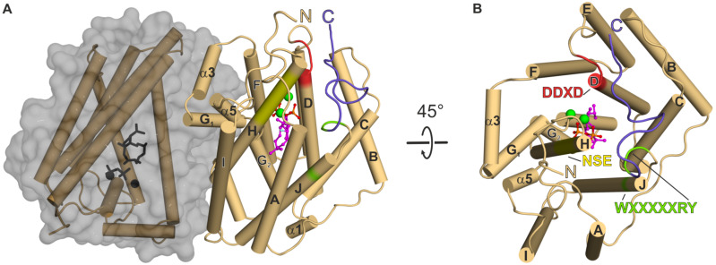
Figure 2.
The bacterial diterpene synthase CotB2wt·Mg2+3·F-Dola in the closed, active conformation (PDB-ID 6GGI; [37]). (A) The two monomers of CotB2 are shown in cartoon representation with the α-helices drawn as cylinders and colored in light brown. One monomer of CotB2 is shown in gray surface representation. The location of the aspartate-rich motif is indicated in red and the NSE-motif is marked in yellow. The WXXXXXRY motif is indicated in light-green. The last 12 C-terminal residues of the lid are drawn in violet. The three Mg2+ ions are represented as green spheres and the bound intermediate F-Dola is shown in magenta. The cleaved diphosphate is shown in orange. (B) View of panel A rotated by 45°. For clarity, one monomer has been removed. The view is into the active site of CotB2.

