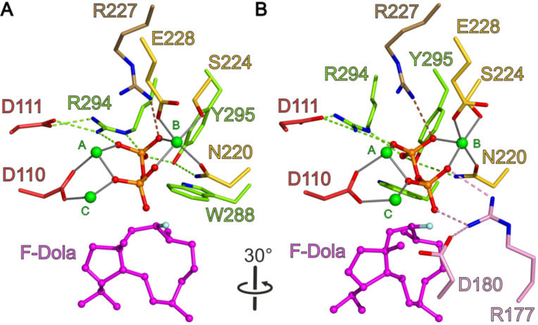
Figure 5.
View into the active site of CotB2wt·Mg2+3·F-Dola [37]. Identical view as in Figure 4. (A) The bound F-Dola reaction intermediate is shown in magenta with the fluorine atom colored in light blue, and Mg2+-ions are shown in green. Residues of the DDXD motif are shown in red and residues of the NSE motif in yellow. The conserved residues of the WXXXXXRY motif are drawn in light green and the R227 that interacts with diphosphate moiety in light brown. The cleaved diphosphate is shown in orange. Hydrogen bonds and salt bridges are indicated by dashed lines in the same color-coding as the involved motifs. Gray lines represent the coordination sphere of the Mg2+-ions. For clarity water molecules have been omitted. (B) View in panel A rotated by 30°. In addition to the motifs shown in panel A, the pyrophosphate sensor motif is depicted in pink.

