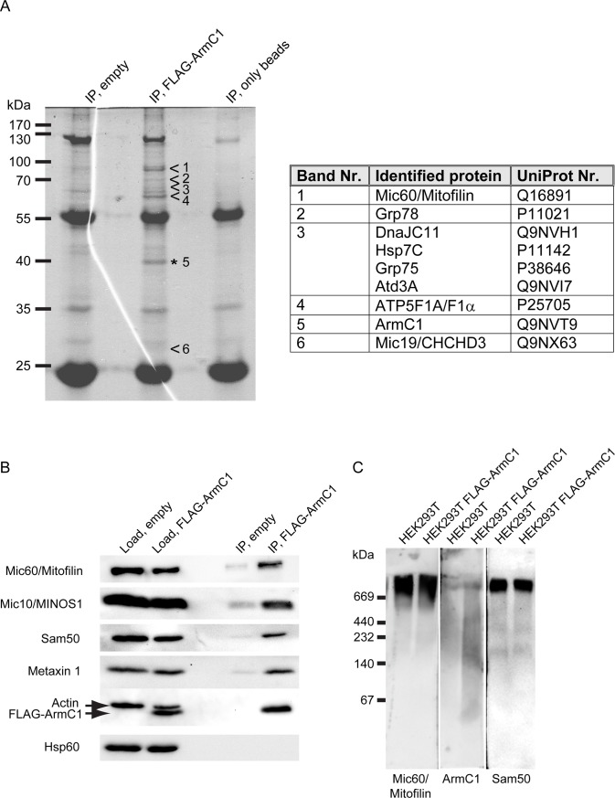
Fig 4. Immunoprecipitation and BN-PAGE analysis of ArmC1.
(A) Mitochondria were isolated from HEK293T cells transfected with an empty pCDNA3 plasmid or with the FLAG-ArmC1 pCDNA3 construct, lysed in the buffer containing 0.5% digitonin and 1 mM PMSF, and incubated with Anti-FLAG M2 affinity gel. Eluted proteins were separated using SDS-PAGE and after colloidal coomassie G-250 staining specific bands were excised from the FLAG-ArmC1 sample and analyzed by mass spectrometry in parallel with the respective regions of the gel from the control empty vector sample. Proteins detected in the FLAG-ArmC1 but not in the control sample are listed in the adjacent table. (B) Immunoprecipitation was performed as in A and samples were analyzed by SDS-PAGE and western blot, using antibodies against Mic60/Mitofilin, Mic10/MINOS1, Sam50, Metaxin 1, Actin, FLAG and Hsp60. (C) Mitochondria were isolated from non-transfected HEK293T cells and cells where FLAG-ArmC1 has been expressed with the help of transient transfection of the FLAG-ArmC1 pCDNA3 plasmid. After solubilization with 1% digitonin buffer, samples were analyzed by BN-PAGE and western blot, using antibodies against Mic60/Mitofilin, ArmC1 and Sam50. MINOS1—mitochondrial inner membrane organizing system 1, Sam50—sorting and assembly machinery 50, Hsp60 –heat shock protein 60.

