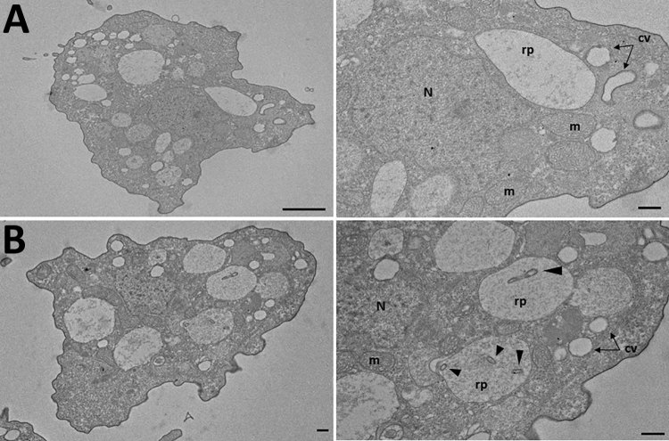
Figure 1.
Transmission electron micrograph of amebae isolated from the home hot tub of a an immunocompromised 3-year-old girl with legionellosis before and after co-culture with Legionella pneumophila, Calgary, Alberta, Canada. A) Trophozoites of Vermamoeba vermiformis before co-culture. Note the absence of intracellular bacteria in the replicative phagosome. B) V. vermiformis replicative phagosome containing L. pneumophila serogroup 6 after 6 h of co-culture. Arrows indicate L. pneumophila contained within replicative phagosomes. Scale bars in left panels indicate 2 μm; scale bars in right panels indicate 500 nm. cv, contractile vacuoles; m, mitochondria; N, nucleus; rp, replicative phagosome.

