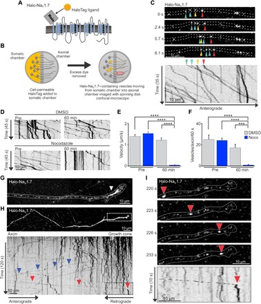
Fig. 2. Anterograde Halo-NaV1.7 vesicles accumulate at the distal axon.

(A) Diagram of the Halo-NaV1.7 construct showing addition of the β4 transmembrane and extracellular location of the HaloTag enzyme. (B) Schematic of the OPAL imaging technique used to visualize anterograde vesicles containing NaV1.7. DRG neurons expressing Halo-NaV1.7 were cultured in MFCs for 7 to 10 days. Immediately prior to imaging, JF549-Halo was added to the somatic chamber for 15 min. After removal of excess dye, axons within the axonal chamber were imaged using spinning disk microscopy to visualize anterograde vesicles traveling from the somatic chamber into the axonal chamber. (C) Representative axon in the axonal chamber labeled as described in (B). Colored arrowheads show locations of the identified vesicles over time. The corresponding kymograph of the Halo-NaV1.7 vesicles was created from a time-lapse image sequence taken at 3.3 frames/s. It shows vesicle position (x axis) as a function of time (y axis). Negative slopes on the kymograph represent movement in the anterograde direction, while vertical lines represent stationary vesicles. (D) Kymographs generated from an axon before and 60 min after either dimethyl sulfoxide (DMSO) or Noco treatment. (E) Average vesicle velocities before and after DMSO or Noco treatment. (F) The number of vesicles with a velocity >0.1 μm/s within a 30-μm segment of axon per 60 s. ****P < 0.0001, ***P < 0.001, two-way ANOVA with Tukey’s multiple comparisons test. (G) Compressed z-stack of an axon terminal in the axonal chamber of the MFC at 1 hour after somatic labeling, demonstrating the accumulation of vesicles near the distal end of the axon (white arrowhead). (H) Image sequence of Halo-NaV1.7 compressed over time to allow visualization of the distal axon. The kymograph of a line scan through this axon shows the behavior of vesicles within this region over time. The vertical lines demonstrate that the vesicles that have accumulated within the distal axon are generally stationary. A small number of channels leave the distal axon as retrograde vesicles (blue arrowheads). Anterograde vesicles move along the axon until reaching the distal axon where they become immobilized (red arrowhead in the black box that corresponds to the region delineated by the white box in the top panel). (I) Time series of a vesicle destined for the distal axon corresponding to the region indicated by the white box in (H), as well as the kymograph region outlined by the back box (H). The red arrowheads follow the position of the vesicle over time as it travels along the axon and then stops at the neck of the distal axon. The vesicle behavior over time is illustrated by the corresponding kymograph with the red arrowhead, denoting where it transitions from mobile to stationary.
