
Figure 2. MTAP deletion promotes tumorigenesis and alters gene expression in GBM cells.
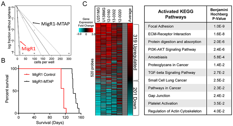
(A) Restoration of MTAP in patient derived GBM cells (13-0302) results in reduced sphere forming capacity, MigR1-Control stem cell frequency is 1/3.68 cells (95% Confidence Interval: 1/2.83 to 1/4.84), MigR1-MTAP stem cells frequency is 1/44.18 cells (95% Confidence Interval: 1/32.91 to 1/59.36), Chi square = 138, P value = 5.76×10⁻³², analyzed using Extreme Limiting Dilution Analysis (ELDA). (B) Restoration of MTAP expression in MTAP-null patient-derived GBM cells (13-0302) resulted in prolonged survival (140 vs 116 days median survival) after intracranial transplantation into immunocompromised (NSG) mice (n=9 animals per arm, Wilcoxon test P value < 1×10⁻⁴). (C) Heatmap showing relative gene expression fold change in MTAP-null cells compared with MTAP-expressing cells of each of the indicated isogenic cell pairs. Included are 520 genes with significantly different expression across all 5 isogenic cell pairs. Right: DAVID analysis of upregulated genes (319 probes) in MTAP-null cells shows significant enrichment of numerous activated KEGG pathways.