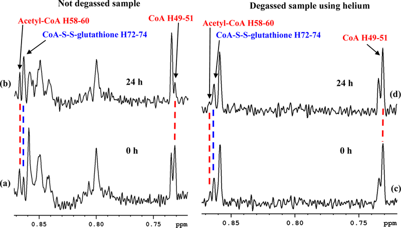
Figure 5.
Portions of 800 MHz 1H NMR spectra of a typical mouse heart tissue extract without degassing: (a) obtained immediately after sample preparation; and (b) obtained 24 h after preparation of the sample. Note, while the acetyl-CoA level is unaltered, the CoA level reduced drastically with a concomitant increase of the CoA-S-S-glutathione level 24 h after sample preparation. Portions of spectra of a typical mouse heart tissue extract with degassing of both the NMR tube and solvent using helium gas: (c) obtained immediately after sample preparation; and (d) obtained 24 h after preparation. Note, the levels of the CoA, acetyl-CoA, and CoA-S-S-glutathione are unaltered even 24 h after the sample preparation, which indicates no oxidation of CoA. Tissue samples used for (a-b) and (c-d) were from different mouse. Peaks around 0.80 and 0.85 ppm observed in (a-b) are unidentified and need to be investigated in future. Peak labels correspond to the hydrogen atom labeling based on the recently developed ALATIS, which creates a unique and atom-specific InChI labels (Figure S3).38

