
Figure 5.
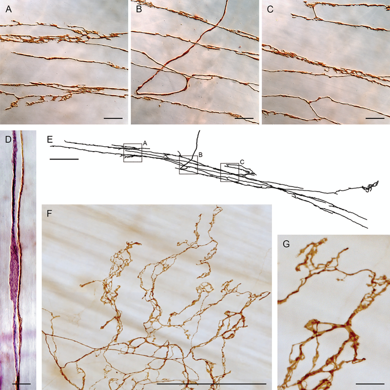
Intramuscular arrays (IMAs), in contrast to vagal preganglionic efferents and IGLE afferents, directly innervate the muscle layers and form arrays of branches that typically run with the ICC network found in the smooth muscle layers. Panels A, B, and C are photomicrographs of branches of the IMA digitized with Neurolucida in panel E. Panel D illustrates how IMA branches (dextran-biotin labeled brown fiber) course with ICCs (elongated purple cell—Vector® VIP labeled for cKit antibody) in the muscle layers. Panel F illustrates the specialized “web ending” variant of IMAs seen near the distal antral/pyloric insertion of sling muscle fibers. Panel G is a higher power image of the IMA apparatus and its contacts or varicosities illustrated in panel F. Scale bars = 20 μm (panels A−C), 10 μm (panel D), 250 μm (panels E and F), and 25 μm (panel G). Panels A–E reproduced by permission of Elsevier from Powley and Phillips.33