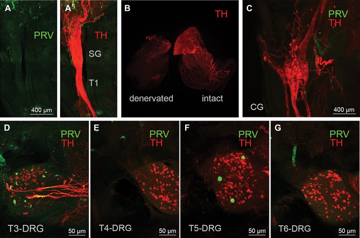
Figure 4.

Controls for nonspecific labeling. (A and B) Light sheet microscope images of the fused right stellate/T1 ganglion showing the absence of PRV‐labeled neurons (green) in mouse with PRV injections into surgically denervated right iBAT pad. (A’) Same ganglion with TH fluorescence (red) to show the outline of the ganglion. The sympathetic innervation (TH labeling) is degraded in the denervated iBAT pad, in contrast to the intact iBAT pad (B). (C) Light sheet microscope image of a mouse with successful PRV labeling in sympathetic chain ganglia shows a complete absence of PRV labeling in the celiac ganglion (CG). (D–G) Confocal microscope images of a mouse with successful PRV labeling in sympathetic chain ganglia show sparse PRV labeling (green dots) in T3‐DRG (D), T4‐DRG (E), T5‐DRG (F), and T6‐DRG (G). TH, tyrosine hydroxylase; PRV, pseudorabies virus; SG, stellate ganglion; DRG, dorsal root ganglion; T1−T7, ganglia for thoracic levels 1−7.
