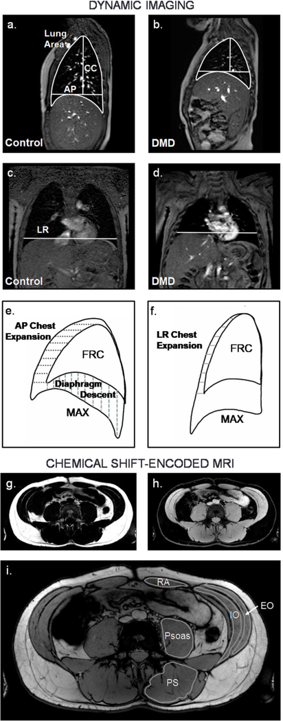
Fig 1. Representative dynamic MRIs, analysis parameters, and chemical shift-encoded MRIs.
Dynamic imaging was performed in the sagittal plane (a-b) and coronal plane (c-d) (Control = 12.8 yrs old, individual with DMD = 12.3 yrs old). Lung size measurements included sagittal plane lung area, anteriorposterior (AP) chest diameter measured at the level of the top of the diaphragm, craniocaudal (CC) length measured from the apex of the lung to the AP chest diameter line, and left-right (LR) chest width measured at the level of the top of the right diaphragm dome. e) Diaphragm movement after a maximal inspiration or expiration was quantified in the sagittal image, and chest movement was quantified in the sagittal and f) coronal images. CSE MRIs were acquired at the chest and abdomen. They were reconstructed to produce fat g) and water h) images, and FF was quantified for the abdominal muscles indicated in the CSE out-of-phase image in i). FRC = functional residual capacity, MAX = maximal inspiration, RA = rectus abdominis, EO = external oblique, IO = internal oblique, PS = paraspinals, CSE = chemical shift-encoded, FF = fat fraction

