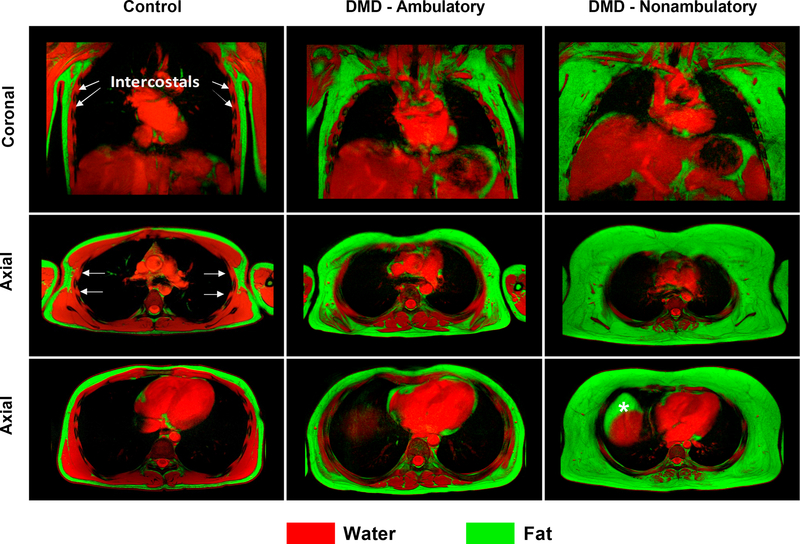
Fig 5. Fat-water fusion images of the chest.
Fat (green) and water (red) images of the chest were acquired in the coronal and axial planes and overlaid to produce fusion images for a 17 yr old unaffected control, an ambulatory 12 yr old with DMD, and a nonambulatory 12 yr old with DMD. In the individuals with DMD, fatty infiltration is visible in the muscles of the chest. The serratus anterior, which is visible overlying the ribs in the coronal image of the control participant is completely infiltrated with fat in the nonambulatory individual with DMD. However, the intercostal muscles, which are accessory respiratory muscles located between the ribs and beneath the serratus anterior, demonstrate less involvement than the other chest muscles. (Note: In the nonambulatory participant, a pocket of fatty tissue, denoted by an asterisk, is visible anterior to the liver and represents true fat rather than a fat-water switching artifact)

