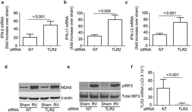
Figure 2.
TLR2-regulated RV-induced IFNs is not due to dysregulation in MDA5 signaling pathway. BEAS-2B cells were either transfected with TLR2 or non-targeting (NT) siRNA, infected with sham or RV and incubated for 24 h and mRNA expression of IFNs was assessed by RT-qPCR (a – c). Data was normalized to G3PDH and expressed as fold change over sham-infected cells. Similarly infected cultures were lysed in RIPA buffer after 4h incubation, and cell lysates containing equal amounts of total protein were subjected to Western blot analysis with antibodies to MDA5 and β-actin (d) or phospho- and total IRF3 (e). Images are representative of 3 independent experiments. Total RNA from NT or TLR2 siRNA-transfected cells was subjected to RT-qPCR to determine the expression of TLR2 and the data normalized to G3PDH (f). Data in a – c, and f represent average ± SEM calculated from three independent experiments (t test).

