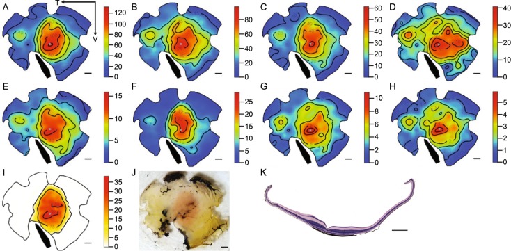
Figure 3.
Retinal topography and histology of Empidonax flycatchers. (A) All cones, (B) all traditional cones, (C) all single cones, (D) P-type oil droplets, (E) R-type oil droplets, (F) Y-type oil droplets, (G) C-type oil droplets, (H) T-type oil droplets, (I) MMOD-complex photoreceptors, (J) trans-illuminated photograph of the retinal wholemount with the orange central area of MMOD-complex photoreceptors clearly visible, (K) cross section showing the fovea (invagination in the retinal tissue) and retinal thickening corresponding to the MMOD-complex photoreceptor region. All numbers are X x103, all scale bars are 1 mm, the arrows in panel A indicate the temporal (T) and ventral (V) directions, gray dots indicate the position of the fovea, and black bands indicate the position of the pecten.

