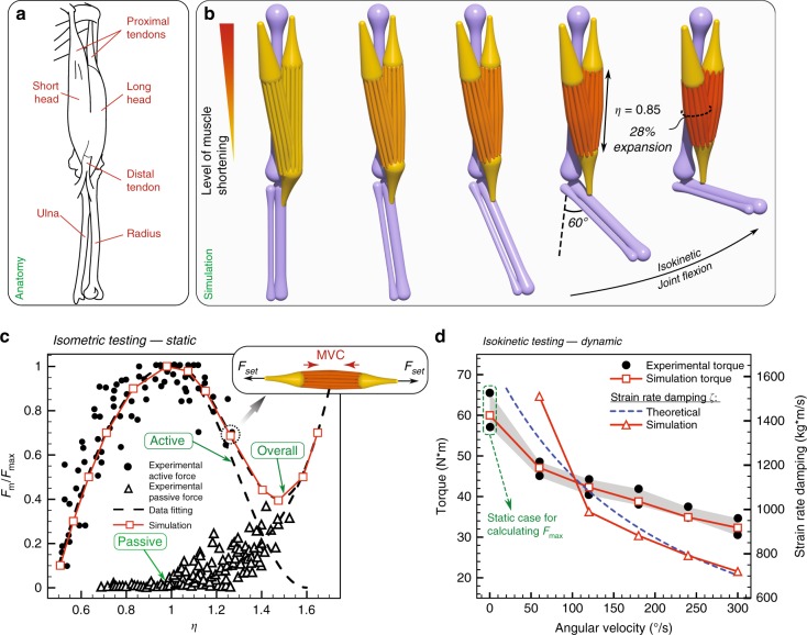
Fig. 1.
Human elbow actuation. a Elbow anatomy. b Simulation of an elbow composed of three bones (humerus, ulna and radius) and two heads of biceps (short and long head) performing a complete flexion. c Experimental data60 and simulations for active and passive force normalized with peak force during the isometric exercise ( mimics the resistance encountered by the muscle and results in its equilibrium length ). d Experimental61 and simulation torque measurements of the elbow joint (angled at 60°) performing maximal isokinetic concentric flexions at different angular velocities along with the corresponding overall muscle strain rate damping . The numerically determined (see Supplementary Note 2) are then compared with theoretical estimates based on the Hill model62

