Fig. 2.
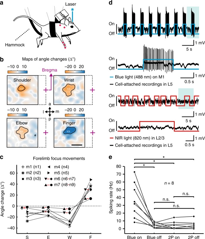
Optogenetic mapping and electrophysiology during two-photon laser scanning. a Motor mapping setup with an anesthetized mouse hanging in a hammock, limbs dangling free. Blue laser light randomly scanned across M1 evoked forelimb joint angle movements that were monitored with a camera. b Maps of evoked joint angle changes for an example mouse. Negative values correspond to flexion (orange, retroversion in the shoulder joint), positive values to extension (blue, anteversion in the shoulder joint). Laser-stimulation elicited responses in shoulder (area 1.12 ± 0.19 mm2), elbow (1.29 ± 0.27 mm2), wrist (1.16 ± 0.32 mm2), and finger-base joints (1.03 ± 0.13 mm2; thresholded at 50% of maximal response in each joint, black contour; mean ± s.e.m. in each case). The superimposed white rectangles indicate the selected forelimb focus area for subsequent calcium imaging (purple cross = bregma, dashed lines indicate the affiliation to the respective map; a = anterior; p = posterior; m = medial; l = lateral). Scale bar 1 mm. c Movement amplitude of angle changes in shoulder (S), elbow (E), wrist (W) and finger-base joints (F) in the seven mice when optogenetic stimulation is applied in the forelimb focus. d Upper part: Cell-attached recording of a ChR2-expressing L5 neuron in M1 during repetitive application of blue 488-nm light. The evoked spiking pattern during one stimulation period (shaded area) is shown on expanded time scale below. Lower part: Cell-attached recording of the same L5 neuron during two-photon excitation laser scanning with near-infrared (NIR) light in L2/3, equivalent to the conditions used for L2/3 calcium imaging. The expanded view of one stimulation period below demonstrates the lack of two-photon excited spikes and extracellular voltage changes. e Pooled data for similar recordings in eight L5 neurons, indicated by black dots (neurons were not recorded from the same ChR2 mice that are shown in (c)). Whereas blue light stimulation induced strong spiking of L5 neurons, laser scanning in L2/3 with two-photon (2P) excitation light of 820-nm wavelength did not induce any detectable changes in the spiking rate of L5 neurons. Asterisks indicate significant differences with P < 0.05, n.s. (non-significant) means P > 0.05 (paired t-test; P-values HB-adjusted)

