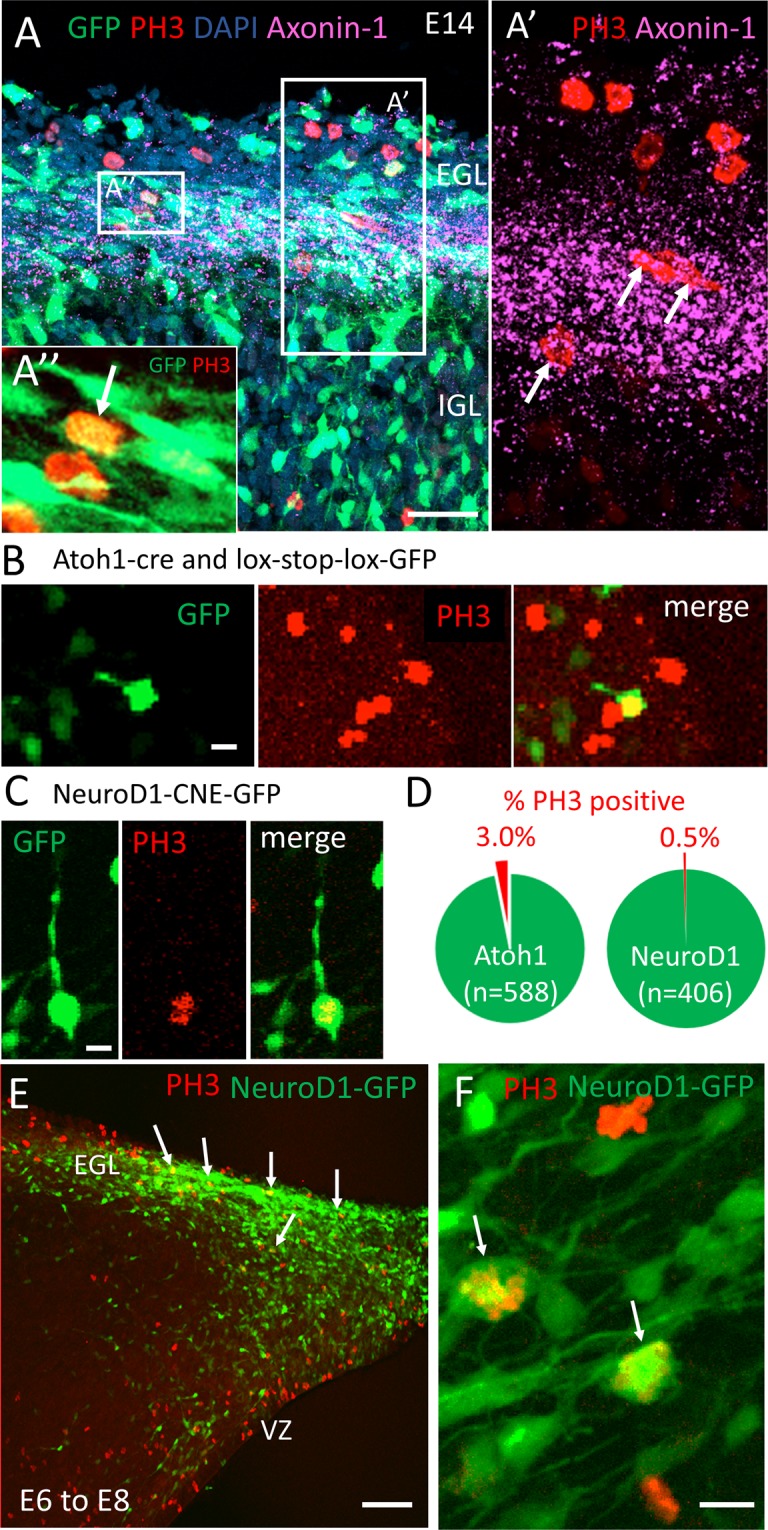
Figure 5.

Mitotically active granule cell precursors can express proteins associated with differentiation such as TAG-1 (Axonin-1) and NeuroD1. (A) Cerebellar tissue from chick embryos electroporated with Tol2:GFP at E4 and fixed at E14 was immunostained for PH3 (red) and TAG1/Axonin-1 (magenta). DAPI shown in blue. (A’) Co-localization of mitotic, PH3 expressing cells and Axonin1 in the inner EGL. (A”) A GFP expressing cell expressing PH3 is surrounded by bipolar cells with long processes in the inner half of the EGL. (B,C) E14 cerebellar slices were electroporated with Atoh1-cre and lox-stop-lox-GFP plasmids (B) or NeuroD1-CNE-GFP plasmid (C) and stained for PH3. (D) Co-localization of PH3 and GFP was observed in 3% of cells where GFP was driven by the Atoh1 enhancer, and in 0.5% of cells where GFP was driven by NeuroD1 enhancer element. (E) A plasmid encoding full length NeuroD1-GFP driven by β-actin promoter was electroporated into the rhombic lip of E6 embryos, fixed two days later, at E8, and stained for PH3. A sagittal cut through the cerebellum reveals cells co-expressing NeuroD1-GFP and PH3 in the forming EGL (arrows). VZ = ventricular zone. (F) A higher magnification of the EGL cells co-expressing NeuroD1-GFP and PH3 (arrows). Scale bar = A,E = 50 μm, B,C,F = 10 μm.
