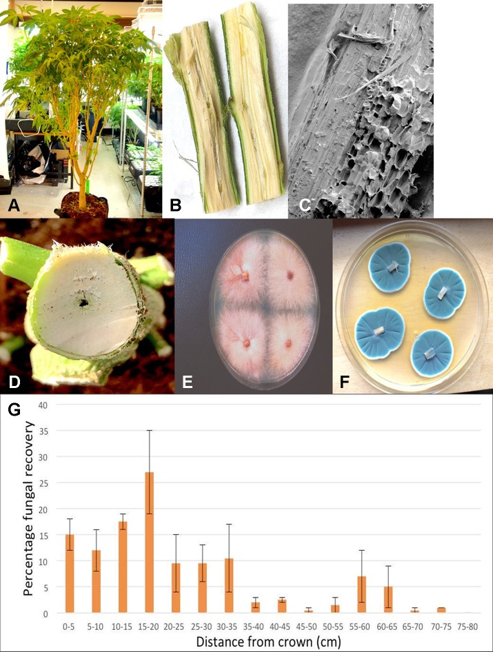
Figure 11.
Recovery of endophytic fungi from cannabis stem tissues. (A) Plant grown indoors in coco substrate and used for sampling studies. (B) Longitudinal section though the main stem near the crown showing the central pith tissue. (C) Scanning electron microscopic image of the pith region showing loosely arranged parenchyma cells (arrow). (D) Young stem higher up the plant showing initial stages of pith development and hollow space. (E) Cross-section through the main stem of a cannabis plant showing the interior of the central pith which has become hollow. (F) Recovery of Fusarium oxysporum from central pith tissues near the crown region of the plant. (G) Recovery of Penicillium chrysogenum from central pith tissues near the crown region of the plant. (H) Frequency of recovery of total fungal species from crown and stem tissues at various distances away from the base of a cannabis plant grown in coco substrate in an indoor environment. Tissues were dissected and surface-sterilized and plated onto PDA+S. Data are from two separate experiments, representing two plants with four replicate dishes at each of 15 sampling distances. Bars show standard errors of the mean.

