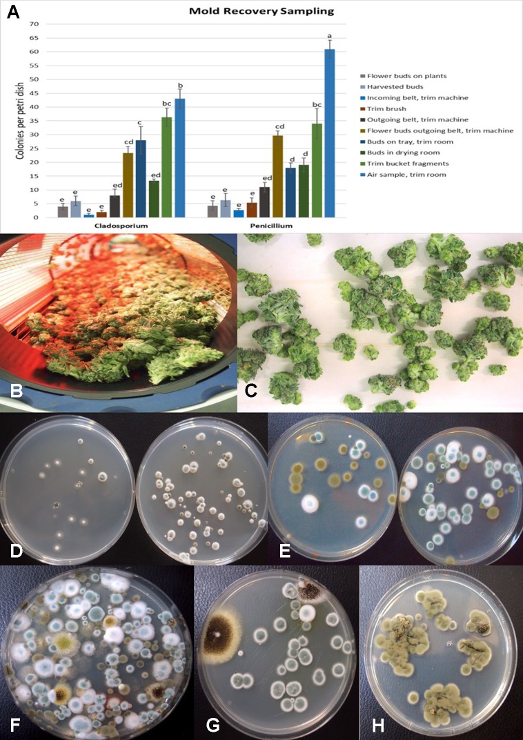
Figure 13.
(A) Recovery of colony-forming units of Cladosporium and Penicillium species on potato dextrose agar at various stages of sampling of cannabis tissues, starting from buds on plants to harvested and mechanically trimmed buds. Swabs were taken of buds entering into a mechanized trim operation at different stages as indicated on the graph. Final samples were taken from trim buckets and air in the trim room, and from dried buds prior to packaging. The data are from three repeated sampling times conducted in two facilities. A minimum of eight replicate petri dishes were used at each sampling stage. Bars show +/− standard errors and were analyzed for significant differences using ANOVA. Means followed by a different letter are different according to Tukey’s HSD test at P = 0.05. (B) Trimmed buds leaving the trim machine. (C) Trimmed buds on the conveyor belt. (D) Recovery of Penicillium species from swabs taken of buds prior to being trimmed (left) compared to buds that had been trimmed (right). (E) Recovery of Penicillium and Cladosporium species from swabs taken of buds prior to being trimmed (left) compared to buds that had been trimmed (right). The number of Penicillium colonies recovered was increased following trimming. (F) Colonies of Penicillium, Aspergillus, and Cladosporium species from air samples collected from within a trim room. Dishes were left exposed for 60 min and taken back to the laboratory to allow for colony development and enumeration. (G) Swabs taken of indoor-grown dried cannabis buds showing growth of Aspergillus niger (black colonies) and Penicillium olsonii (blue-green colonies). (H) Swabs taken of dried field-grown cannabis buds and tissue segments plated on potato dextrose agar showing development of Cladosporium westerdijkieae.

