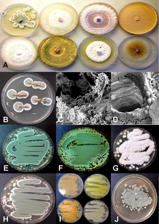
Figure 14.
Colony morphology of endophytic fungi and contaminant fungi recovered from cannabis tissues. All colonies were grown potato dextrose agar. (A) Endophytic fungi recovered from surface-sterilized stems, petioles, and nodal segments after 10 days of growth in culture. From left to right (top row) are Penicillium chrysogenum, Fusarium oxysporum, F. oxysporum, and a Fusarium sp. Bottom row—Trametes (Polyporus) versicolor, Trichoderma harzianum, Simplicillium lanosoniveum, and Chaetomium globosum. (B) Emergence of Penicillium olsonii from stem pieces following surface-sterilization, indicating that internal colonization of tissues had occurred. (C, D) Scanning electron micrographs of dissected pith tissues from cannabis stems showing profuse sporulation of P. olsonii and spores adjacent to parenchyma pith cells. (E–H) Cultures of Penicillium species streaked out from swab transfers made from pure cultures originating from cannabis buds and incubated for 96 h to show colony color development. (E) Penicillium spathulatum. (F) Penicillium citrinum. (G) Penicillium simplicissimum. (H) Penicillium olsonii. (I) The underside of colonies of the same four Penicillium species after growth for 96 h. The unique colors of these species could be used for preliminary identification purposes. (J) Colony of Aspergillus sydowii after 2 weeks of growth originating from cannabis bud tissue.

