
Figure 6.
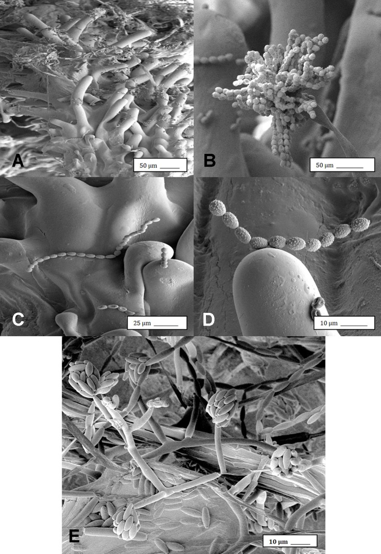
Scanning electron microscopic images of the development of mycelium and spore production by Penicillium olsonii on inoculated cannabis inflorescences. (A) Spores and mycelium on stigmatic hairs (papillae). (B) Conidiophore with chains of conidia characteristic of Penicillium formed on the bud surface. (C, D) Close-up views of spore chains of P. olsonii stuck to stigmatic hairs. (E) Conidiophore and conidia of Fusarium oxysporum on inoculated flower bud.