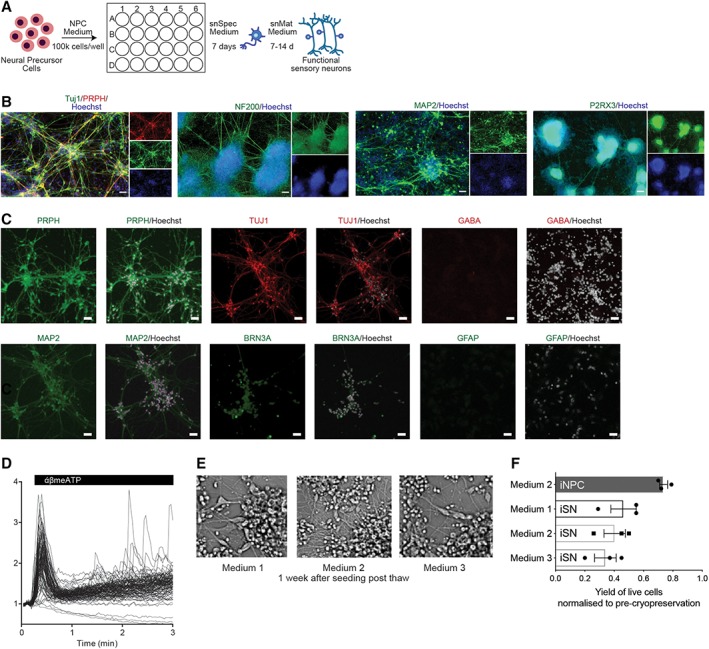
Figure 2.

Sensory neuron differentiation of direct conversion neural precursor cells. (A): Schematic of sensory neural differentiation protocol from peripheral blood‐derived induced neural progenitor cells (iNPCs). (B): Immunofluorescence images of 21 day differentiated induced sensory neurons (iSNs). Cells were fixed and stained for peripheral nervous system (PNS) markers, Tuj1, PRPH, NF200, MAP2, and P2RX3. Nuclei were stained with Hoechst. Scale bar represents 50 μM. (C): Immunostaining of iSNs cultures with PNS specific, CNS specific and glial markers, PRPH, Tuj1, MAP2, GABA, BRN3a, and GFAP. Scale bar represents 50 μM. (D): Calcium trace and distribution of cells in response to 30 μM α,β‐meATP treatment. (E): Phase contrast images of iSNs 1 week post‐thaw for different cryopreservation medium. (F): Optimization of cryopreservation of iNPCs and iSNs. Yield of viable cells after cryopreservation in various medium. Mean ± SD for three independent experiments.
