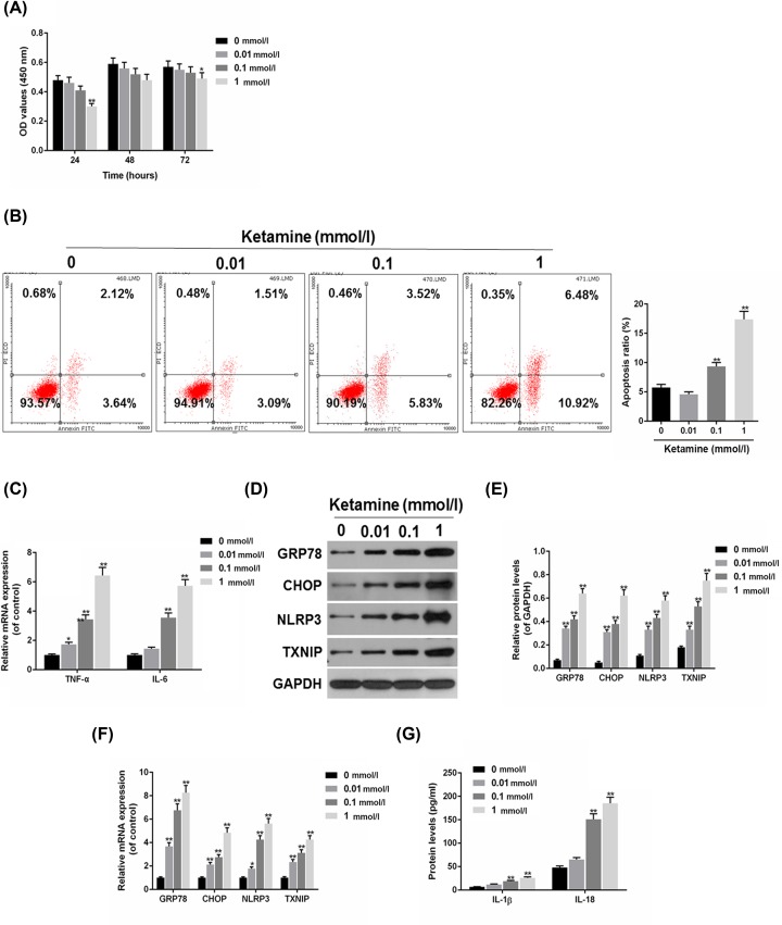
Figure 2. Cell apoptosis and NLRP3/TXNIP were induced by ketamine in SV-HUC-1 cells.
(A) The viability of SV-HUC-1 cells treated with 0.01, 0.1 and 1 mmol/l ketamine for 24, 48 and 72 h were determined by CCK-8. (B) Ketamines at 0.01, 0.1 and 1 mmol/l was used to treat SV-HUC-1 cells apoptosis, which was analyzed by flow cytometry. (C) TNF-α and IL-6 mRNA levels were analyzed by RT-qPCR. (D,E) The protein levels of GRP78, CHOP, NLRP3 and TXNIP were determined (D) and quantified (E) by Western blot. (F) The mRNA levels of GRP78, CHOP, NLRP3 and TXNIP were determined by RT-qPCR. (G) The protein levels of IL-1β and IL-18 were measured by ELISA. *P<0.05, **P<0.01 vs. control.

