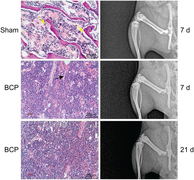
Figure 1.
X-ray scanning and HE staining indicate the successful establishment of mouse models of BCP. Yellow arrows indicate normal trabecular bone structure and the black arrow indicates the destroyed trabecular bone structure by cancer cells on day 7. On day 21, almost all trabecular bone structures were destroyed.
BCP, bone cancer pain; HE, hematoxylin-eosin.

