
Figure 2.
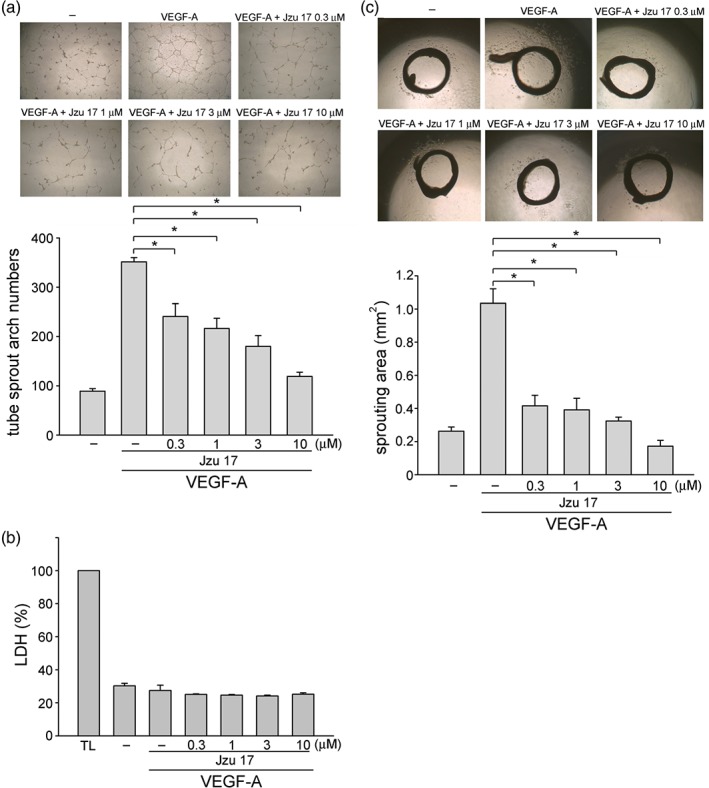
Jzu 17 inhibited VEGF‐A‐induced tube formation in vitro and aorta ring sprouting ex vivo. (a) HUVECs were seeded on Matrigel in the presence of VEGF‐A (200 ng·ml−1) with or without Jzu 17 at indicated concentrations. Cells were photographed under phase contrast after 16 hr as described in Section 2. Bar graphs show compiled data of average sprout arch numbers (n = 6). *P < .05, significantly different from VEGF‐A alone; one‐way ANOVA, with Tukey's post‐hoc test. (b) Cells were stimulated with VEGF‐A (25 ng·ml−1) with or without Jzu 17 at indicated concentrations for 16 hr. Jzu 17's cytotoxicity was determined by LDH assay. Cells were also treated with cell lysis buffer (total lysis, TL) to serve as positive control. Each column represents the mean ± SEM of six independent experiments performed in duplicate. Technical replicates were used to ensure the reliability of single values for each experiment. *P < .05, significantly different from VEGF‐A alone; Kruskal–Wallis test. (c) Rat aortic rings were placed in Matrigel and treated with VEGF‐A (25 ng·ml−1) in the presence or absence of Jzu 17 at indicated concentrations. The effects of Jzu 17 on formation of vessel sprout from various aorta samples was determined on Day 8. Bar graphs show compiled data of average microvessels area (n = 7). *P < .05, significantly different from VEGF‐A alone; one‐way ANOVA, with Tukey's post‐hoc test